What Is a Sonogram vs Ultrasound? Function, Assessment & Diagnostic Use – Safe, Radiation-Free Imaging in 30 Minutes - MaNaDr Medical Notes - Manadr
What Is a Sonogram vs Ultrasound? Function, Assessment & Diagnostic Use – Safe, Radiation-Free Imaging in 30 Minutes
MaNaDr2024-03-21
Ultrasound, also widely known as sonography or a sonogram, is a non-invasive medical imaging technique that plays a crucial role in modern diagnostics. Unlike X-rays or CT scans, it operates without any ionizing radiation, making it a remarkably safe tool for visualizing internal body structures. From monitoring pregnancies to assessing organ health and guiding precise medical procedures, ultrasound imaging offers unique insights into the body’s condition.
This comprehensive guide will delve into what is an ultrasound and its interchangeable terms, explore how do ultrasounds work and what can an ultrasound detect, detail the broad range of diseases it can help diagnose, confirm its safety profile, and explain what to expect during an ultrasound procedure, often completed in just 30 minutes.
1. What Exactly Is an Ultrasound (Sonogram)?
To truly grasp the capabilities of this technology, it’s essential to understand its fundamental definition and the precise meaning behind its various names.
1.1. Defining Ultrasound and Sonography
Ultrasound is a medical imaging technique that uses high-frequency sound waves to create live images from inside the body. For those asking, “what is an ultrasound” or “what is ultrasound,” it’s a diagnostic tool that provides a real-time view of soft tissues, organs, and blood flow. It’s also often referred to as ultrasonography or an ultrasound scan.
The ultrasound definition highlights its non-invasive nature and its ability to capture dynamic images of internal structures without relying on surgery or radiation. This ultrasound test is widely used across many medical specialties.
1.2. Ultrasound vs. Sonogram: Are They Different?
The terms “ultrasound” and “sonogram” are frequently used interchangeably, leading to questions like “what is a sonogram vs ultrasound?” or “whats a sonogram?” In most clinical contexts, they refer to the same procedure.
Ultrasound: This term refers to the technology itself—the use of high-frequency sound waves for diagnostic purposes. It’s the overarching method of ultrasound imaging.
Sonogram: This term specifically refers to the actual image or visual record produced by the ultrasound scan. So, when you receive a printout of an ultrasound image, it’s technically a sonogram. The process of performing the scan is sonography.
Therefore, “what is a sonogram” is essentially asking about the visual output of an ultrasound procedure. The healthcare professional who performs the scan is called a sonographer.
1.3. How Ultrasound Differs from Other Imaging (e.g., X-ray, CT)
Ultrasound stands apart from many other common medical imaging techniques due to its unique principles:
Radiation-Free: Unlike X-rays, CT (computed tomography) scans, or PET (positron emission tomography) scans, ultrasound does not use ionizing radiation. This makes it particularly safe for repeated use, and for sensitive populations like pregnant individuals and children. This directly addresses “does ultrasound use radiation?“
Soft Tissue Visualization: While X-rays excel at bone imaging, ultrasound is superior for visualizing soft tissues such as organs, muscles, tendons, and blood vessels.
Real-Time Imaging: A key advantage of ultrasound is its ability to provide dynamic, real-time images. This allows healthcare providers to observe organs in motion, assess blood flow, and guide procedures in real-time. This is why it’s also called ultrasonography.
1.4. Expert Insight: Beyond Pregnancy
Expert Insight: A common myth is that “Ultrasound is primarily, or even exclusively, used for monitoring pregnancy and seeing babies.” While its role in obstetrics is undeniably vital, leading radiologists and medical professionals emphasize the crucial fact that ultrasound is an incredibly versatile diagnostic tool utilized across almost every medical specialty. Its applications range from cardiology (for echocardiograms), emergency medicine (for rapid internal bleeding assessment), and guiding biopsies to assessing musculoskeletal injuries (tendons, muscles), evaluating abdominal organs, and examining the thyroid and breasts. This broad utility makes it a foundational and frequently used imaging modality far beyond prenatal care.
2. How Do Ultrasounds Work and What Can They Show?
Understanding the fundamental principles behind ultrasound technology clarifies its unique capabilities in diagnostic medicine. The way ultrasound generates images is based on sound waves, allowing it to reveal detailed information about internal structures and even dynamic processes.
2.1. The Science Behind Ultrasound Technology
The operation of ultrasound relies on the physics of sound waves:
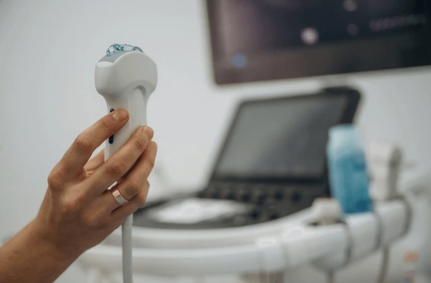
Transducer’s Role: A small, handheld device called a transducer (or probe) is placed directly on the skin, often with a gel applied to ensure good contact. The transducer contains piezoelectric crystals.
Sound Wave Emission: When electricity passes through these crystals, they vibrate rapidly, producing high-frequency sound waves (ultrasound waves) that are beyond the range of human hearing. These waves travel into the body. This explains “how does ultrasound work” at a fundamental level.
Echo Reception: As these sound waves encounter various organs, tissues, and fluids within the body, they bounce back (echo) to the transducer. The transducer then converts these echoes back into electrical signals. This is essentially “how does a sonogram work.”
Computer Processing: A sophisticated computer processes these electrical signals, interpreting the strength, direction, and time it took for the echoes to return. Based on this data, the computer constructs a live, two-dimensional image (or even 3D/4D images for specialized ultrasounds) on a screen. The different shades of gray on the ultrasound image represent varying tissue densities.
This real-time processing of sound waves allows for dynamic assessment, showcasing not just static structures but also movement and flow.
2.2. What Can an Ultrasound Show? (Applications)
Ultrasound excels at visualizing specific types of body structures and dynamic processes:
Soft Tissues and Organs: It provides excellent images of organs like the liver, kidneys, gallbladder, pancreas, spleen, bladder, uterus, and ovaries. It can identify the presence of masses, cysts, or fluid collections within these organs. This directly answers “what does an ultrasound show” in terms of anatomical structures.
Blood Vessels and Blood Flow (Doppler Ultrasound): Specialized ultrasound technology known as Doppler ultrasound can measure the direction and speed of blood flow within blood vessels. This is invaluable for detecting blockages (e.g., deep vein thrombosis – DVT), narrowing (stenosis), or aneurysms.
Muscles, Tendons, and Ligaments:Ultrasound imaging is increasingly used to assess musculoskeletal injuries such as tendon tears, muscle strains, and ligament damage, and to identify fluid collections or inflammation around joints.
Real-time Movement: The dynamic nature of ultrasound allows healthcare providers to observe organs in motion, such as a beating heart (echocardiogram) or the movement of a fetus during pregnancy. This real-time capability is a major advantage for “what can an ultrasound detect” that static imaging cannot.
2.3. Limitations of Ultrasound
Despite its versatility, ultrasound does have certain limitations:
Cannot Penetrate Bone or Air: Sound waves are effectively blocked by bone and scattered by air. This means ultrasound is not ideal for imaging structures completely surrounded by bone (like the brain inside the skull in adults) or organs filled with gas (like the lungs or large parts of the bowel).
Operator Dependent: The quality of the ultrasound scan is highly dependent on the skill and experience of the sonographer performing the exam. Acquiring clear and accurate images requires significant expertise.
3. What Diseases Can Ultrasounds Detect and Diagnose?
The widespread adoption of ultrasound in medicine is a testament to its versatility in detecting and diagnosing a broad spectrum of conditions across various body systems. Its diagnostic ultrasound capabilities make it an invaluable tool.
3.1. Broad Diagnostic Use Cases
Ultrasound has become an indispensable tool across numerous medical specialties for ultrasound assessment and ultrasound diagnosis. Its non-invasive nature and real-time capabilities make it a preferred initial imaging modality for many concerns.
3.2. Common Applications by Body System
Here are some of the most common applications of ultrasound and what diseases can be detected by ultrasound:
Gallbladder: Diagnosing gallstones and inflammation (cholecystitis).
Kidneys: Identifying kidney stones, cysts, tumors, and assessing kidney function (e.g., hydronephrosis).
Pancreas and Spleen: Assessing for inflammation, cysts, or masses.
Bladder: Evaluating bladder stones, tumors, or incomplete emptying.
Cardiovascular System:
Echocardiogram: Assessing heart structure, function, valve issues, and blood flow within the heart.
Vascular Ultrasound: Detecting blood clots (e.g., deep vein thrombosis – DVT), narrowing of arteries (e.g., carotid artery stenosis), or aneurysms in various blood vessels.
Musculoskeletal System:
Tendons and Muscles: Diagnosing tears, strains, tendonitis, and other soft tissue injuries.
Joints: Assessing for fluid accumulation, inflammation, or certain ligament injuries.
Thyroid and Neck:
Identifying thyroid nodules, goiter, and assessing lymph nodes in the neck.
Breast:
As an adjunct to mammography, ultrasound helps evaluate breast lumps, cysts, and other abnormalities, especially in dense breast tissue.
Urological System:
Assessing the prostate for enlargement or abnormalities, and evaluating the testes for masses or inflammation.
Emergency Medicine:
Rapid assessment (e.g., FAST exam in trauma) to detect internal bleeding or fluid accumulation in the abdomen or chest.
3.3. Guiding Procedures
Beyond diagnosis, ultrasound is widely used to guide various medical procedures, enhancing precision and safety:
Biopsies: Guiding needles to obtain tissue samples from suspicious masses (e.g., in the breast, liver, thyroid) for pathology analysis.
Fluid Drainage: Assisting in the drainage of fluid collections (e.g., abscesses, cysts).
Injections: Guiding injections into joints or soft tissues for pain relief.
4. Is Ultrasound Imaging Safe and Does It Use Radiation?
One of the most significant advantages of ultrasound imaging is its remarkable safety profile, particularly its distinction from other common imaging modalities regarding radiation.
4.1. The Safety Profile of Ultrasound
Ultrasound is generally considered one of the safest medical imaging techniques available. Its widespread use in prenatal care, for example, is a testament to its established safety record. It’s a non-invasive procedure that rarely causes discomfort or adverse effects when performed correctly by trained professionals.
4.2. Does Ultrasound Use Radiation?
A crucial fact to understand about ultrasound is that it does NOT use ionizing radiation. For those asking, “does ultrasound use radiation?” or “is ultrasounds radiation?” the answer is definitively no.
Sound Waves, Not Radiation: Unlike X-rays, CT scans, or PET scans, which utilize ionizing radiation (a form of energy that can damage DNA), ultrasound relies entirely on high-frequency sound waves. These sound waves are generated by a transducer and travel through the body, bouncing off structures to create an image.
Radiation-Free Imaging: This characteristic makes ultrasound particularly valuable for repeated examinations, as there is no cumulative radiation exposure. It is the imaging modality of choice for pregnant individuals and children, who are more sensitive to radiation exposure, offering a safe way to monitor health without associated risks.
4.3. Potential Risks and Considerations
While generally very safe, some theoretical considerations exist, although these are minimal in diagnostic use:
Thermal Effects: High-intensity ultrasound waves can potentially generate a small amount of heat in tissues. However, diagnostic ultrasound devices operate at intensities far below levels that would cause significant tissue heating, and operators are trained to minimize thermal effects.
Cavitation: Extremely high-intensity ultrasound could theoretically create tiny gas bubbles in tissues (cavitation). Again, diagnostic ultrasound operates at levels well below this threshold, and there is no evidence of cavitation occurring in human diagnostic procedures.
The benefits of diagnostic ultrasound in providing essential medical information far outweigh these minimal and largely theoretical risks. The American Institute of Ultrasound in Medicine (AIUM) regularly reviews and updates its safety guidelines to ensure the continued safe use of ultrasound.
4.4. Expert Insight: Not All Imaging is Equal
Expert Insight: A common myth is that “all medical imaging procedures carry radiation risks, so I should limit them.” However, leading radiology and public health organizations emphasize the crucial fact that imaging modalities differ significantly in their use of energy. Unlike X-rays, CT scans, or PET scans which utilize ionizing radiation, ultrasound relies entirely on high-frequency sound waves. This makes it a radiation-free imaging modality, particularly valuable for pregnant individuals, children, and those requiring frequent monitoring, as it eliminates any concerns about cumulative radiation exposure. Understanding this distinction is key to making informed decisions about diagnostic testing.
5. What to Expect During an Ultrasound Procedure (Time & Results)?
Knowing what to expect during an ultrasound procedure can help alleviate anxiety and ensure you are well-prepared for your diagnostic exam. The process is generally straightforward, quick, and non-invasive.
5.1. Preparing for Your Ultrasound
Preparation for an ultrasound procedure varies depending on the body part being examined:
Abdominal Ultrasound (e.g., gallbladder, liver, pancreas): You may be asked to fast for several hours beforehand (e.g., 6-8 hours) to ensure certain organs are empty and to reduce gas in the bowel, which can interfere with image quality.
Pelvic Ultrasound (e.g., bladder, uterus, ovaries): You might be asked to drink several glasses of water an hour before the exam and avoid urinating to ensure a full bladder. A full bladder helps visualize pelvic organs more clearly.
Other Ultrasounds (e.g., thyroid, vascular, musculoskeletal): Typically require no special preparation.
Comfortable Clothing: Wear loose, comfortable clothing that allows easy access to the area being examined.
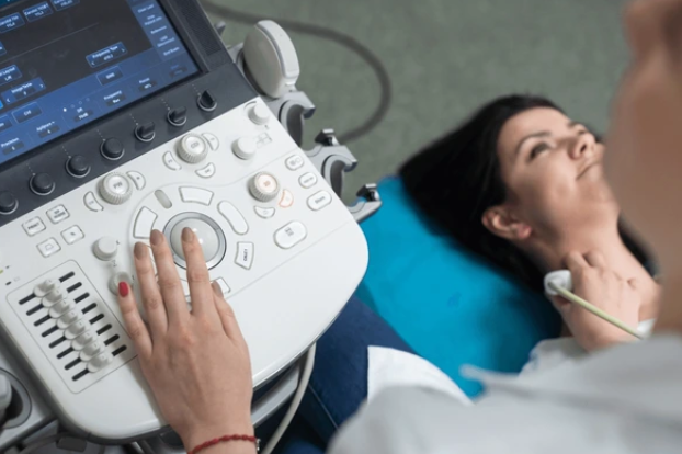
5.2. During the Ultrasound (The Process)
For those wondering “how is ultrasound done?” or “whats ultrasound” like in practice, here’s what typically happens:
Positioning: You will usually lie on an examination table, exposed only in the area being scanned.
Gel Application: A clear, water-based gel will be applied to your skin over the area to be examined. This gel helps the transducer make full contact with your skin, eliminating air pockets that could block the sound waves.
Transducer Movement: A trained sonographer will then press a small, handheld device called a transducer (or probe) firmly against your skin and move it back and forth over the area. You might feel some pressure, but it should not be painful.
Image Capture: The transducer sends and receives sound waves, and a computer converts these echoes into real-time images displayed on a monitor. The sonographer will capture various images and may ask you to hold your breath or change position briefly.
Comfort Level: The procedure is generally painless. You might feel a cool sensation from the gel or slight pressure from the transducer, but no discomfort beyond that.
5.3. How Long Does an Ultrasound Take?
A common question is “how long does an ultrasound take?” or “how long do ultrasounds take?” The duration of an ultrasound procedure can vary significantly depending on the type of exam and the complexity of the findings:
Typical Duration: Most routine ultrasound scans are completed within 15 to 60 minutes. Many, especially focused exams, can be finished in around 30 minutes, as indicated in the article title.
Factors Affecting Time: More complex exams (e.g., detailed fetal anatomy scans, extensive vascular studies) or those where abnormalities are found may take longer.
5.4. Receiving Your Ultrasound Results
After the ultrasound procedure, you won’t typically receive immediate results from the sonographer.
Interpretation by Radiologist: The images captured during the ultrasound are formally interpreted by a radiologist (a doctor specializing in medical imaging) or another specialist. They will analyze the ultrasound image in detail.
Report to Referring Doctor: The radiologist then compiles a comprehensive report of their findings and sends it to the doctor who ordered your ultrasound test.
Timeframe for Results: The time it takes to get your ultrasound results can vary. For urgent findings, results may be available within hours. For routine exams, it might take a few days for your referring doctor to receive the report and discuss the findings with you. It’s best to ask your referring doctor when and how you can expect your results.
If you have questions about preparing for your ultrasound procedure, need help understanding your ultrasound results, or simply want to discuss a condition that might require imaging, MaNaDr offers a convenient platform. You can connect with qualified doctors instantly to get expert advice and guidance from home.
Disclaimer: This article is for informational purposes only and does not constitute medical advice. Always consult with a qualified healthcare professional for diagnosis and treatment of any medical condition.